
免疫学の研究
複雑な動き:リンパ球とその機能
免疫系は、異物の侵入に対する身体の反応を仲介するもので、リンパ球を含む複数の種類の細胞で構成されています。リンパ球は、B細胞(抗体を分泌)、細胞傷害性T細胞、ヘルパーT細胞(サイトカインを分泌)、ナチュラルキラー(NK)細胞から構成されています。多くのサイトカインとシグナル伝達タンパク質が、すべてのリンパ球の適切な制御と機能において複雑な役割を担っています。これらのシグナル伝達経路の特性は、リンパ球の相互作用や免疫系の複雑な性質を理解するために不可欠です。
自然免疫系と適応免疫系の細胞は、協調して病原体の攻撃から体を守るために働いています。抗体は、感染症やワクチン接種などの抗原曝露に対する血清学的反応として発現し、急性および長期的な防御を提供する手段となっています。多くの異なるタイプの免疫細胞は、細胞表面マーカーによって区別されるだけでなく、様々な病原体や刺激に対する多様な細胞応答によって区別されます。
注目のスポットライト:
ワクチンによる細胞性免疫の定量化のためのマルチプレックスサイトカインアッセイのデザイン方法

テキサス州ヒューストンで開催されたxMAP® Connect 2022での、Jeroen Pollet博士による発表
Luminexは、お客様の解析ニーズに合ったマルチプレックスツールを提供し、免疫学研究へのアプローチを簡素化します。
LuminexはビーズベースのxMAP® テクノロジー を提供し、お客様の免疫学研究のニーズを満たします。マルチプレックスイムノアッセイ、遺伝子発現解析、ジェノタイピングなど、必要なツールを取り揃えています。
Flow Cytometry for Immunology
Imaging Flow Cytometry used for Immunology
By combining quantitative image analysis tools with the large sample sizes common to flow cytometry, the Amnis® ImageStream®X Imaging Flow Cytometer uniquely enables multiple applications within the field of immunology, including measurement of nuclear translocation of transcription factors, T:APC interactions and accumulation of proteins at the immune synapse, chemokine-induced shape change, and phagocytosis, just to name a few. Amnis imaging flow cytometers can readily analyze more complex assays, with up to 12 parameters, including visual verification of specific cells in suspension and rare events.
Localization of ADAP to the Immunological Synapse
Ovalbumin-specific accumulation of ADAP at the immune synapse is measured. Without specific peptide ADAP (green) is distributed around the T cell and in the presence of specific peptide ADAP moves to the synapse between the APC and T cell. Quantitation of conjugate formation and representative cells images show that without specific peptide ADAP (green) is distributed around the T cell and in the presence of specific peptide ADAP moves to the synapse between the APC and T cell. See the application note for more details.
Images to be included.
NF-kB Translocation in T:APC Conjugates
Images to be included.
Measurement of NFκB translocation in transgenic T cells which are in contact with antigen presenting cells (APC) and specific peptide. Specific T cell:APC conjugates are identified and translocation of NFκB from the cytoplasm to the nucleus specifically within the T cells is measured in the presence (shown) or absence of peptide. See the application note for more details.
Phagocytosis by Murine Macrophages
Internalization of zymosan (green) by murine RAW cells (orange) identified by immunophenotyping. Phagocytosis is measured as the percentage of cells with internalized zymosan at 15, 30 and 60 minutes at 37 degrees C.
Images to be included.
HIV-Specific Translocation of NFAT
Images to be included.
HIV-specific nuclear localization of NFAT was measured in HIV-experienced T cells from peripheral blood of HIV+ patients. HIVgag peptide specifically stimulates NFAT (green) to move to the nucleus (red) in HIV-tetramer positive cells (orange). The Similarity score correlates DRAQ5 nuclear stain with the NFAT signal. The higher the Similarity score, the more translocation is visualized in the example images from each quadrant.
Conventional Flow Cytometry for Immunology
Guava easycyte™ Systems offer rapid interrogation of a variety of common immunologic assays, with up to 8 parameters of detection. The Guava® Muse® Cell Analyzer brings the essential immunology analyses you perform the most right to your bench top. Obtain rapid single cell analysis of total human CD4, CD8, and B cell counts, relative CD25 expression, and lymphocyte activation. Using optimized assays that eliminate the need for voltage detector setup, modules for each assay automatically calculate absolute cell counts and subpopulation ratios as percentages of total sample.
Our Guava immunology kits have a unique combination of directly conjugated antibodies and/or fluorescent dyes and protein reporters to monitor changes in protein expression and posttranslational modification. The kits also contain complete buffer sets and protocols, and they are fully optimized for “plug-and-play” cellular analysis on Guava and other flow cytometers.
Multiparametric phenotypic analysis of cellular events and/or cell cell phenotypes by flow cytometry allows researchers to study the dynamics of immune signaling in intact cells, and offers many unique capabilities and advantages, such as:
- Distinguishing one subpopulation of cells within a heterogeneous mixture
- Evaluating the impact of activation by antigens
- Detecting suppression of normal immune activation or impact of disease state on phenotypes
- Multiplex detection with high reproducibility
- Optimized antibodies and buffers to remove the need for assay development and optimization
3-part differential capabilities
3-part differential with antibody confirmation (lymphocytes red CD3, monocytes yellow CD14, granulocytes green CD15). Adult human blood stained with CD3PECY5, CD14-PE, and CD15-FITC lysed with the guava® lysing solution and acquired on the guava easyCyte™ instrument. Data displayed shows clear identification of the 3 major white blood cell populations.
Include image
Immunotoxicology
T-cell mediated cytotoxicity plays a critical role in down regulation of the immune response. Moreover, immunosuppressant compounds often mediate their effects through immunotoxicity. We have developed a broad range of kits for evaluation of immunotoxicology through a variety of mechanisms.
Include image
In the example to the right, PBMCs were treated with 80 different cytotoxic compounds for 20 hours. Cells were analyzed with the FlowCellect™ Human T Cell Apoptosis and CD4 T Cell FAS assays followed by microcapillary cytometry. Levels of apoptosis, assessed by Annexin V response, in CD4 and CD8 subsets for multiple compounds are depicted in heat map. The table summarizes compounds that demonstrate significant apoptosis and the % of CD4 and CD8 T cells that underwent apoptosis.
Cells of the Human Immune System
AutoCD4/CD4% (Human CD4 T cell enumeration and percent)
Guava® AutoCD4/CD4% is an application for enumeration of CD4+ Human T cells in whole blood samples. This application is offered on a dedicated use instrument platform. It provides counts of CD4+ T Lymphocytes in whole blood as well as determination of the number of CD4+ T-cells as a percent of Total Lymphocytes.
Include image
Learn More About the Guava® AutoCD4/CD4% Kit
Muse® Human CD4 T Cell Kit
The Muse® Human CD4 T Cell Kit allows you to determine the concentration and percentages of CD4 T cells and lymphocytes in human whole blood or peripheral blood mononuclear cells (PBMCs). Characterizing CD4 T cell counts and percentages is important in evaluating immune status, understanding disease mechanism, in vaccine research and immunotoxicity studies.
Learn more about the Muse® Human CD4 T Cell Kit
Muse® Human CD8 T Cell Kit
The Muse® Human CD8 T Cell Kit is used in laboratory research studies to determine the CD8 T-cell count, total lymphocyte count, and CD 8 T-cell percent of lymphocytes in human whole blood and peripheral mononuclear cell (PBMC) samples. Identification and enumeration of cytotoxic CD8 T-cell populations are useful in understanding immune response and status, and in the research of disease mechanisms, such as autoimmune diseases, and immunodeficiency.
Learn more about the Muse® Human CD8 T Cell Kit
Human B Cell Function
Human B Cell subpopulations are reported to be defective in patients suffering from immuno-deficiency disorders, which are correlated with reductions in numbers of circulating CD19+CD27+ memory B cells. For evaluation of Human B cells, we now offer a variety of options.
Identification of Human Memory B cells by using specific phenotypic CD markers
Include image
Isolation and Identification of Human Memory B cells from human PBMSs. Human memory B cells are phenotypically identified by using specific human CD markers: Human cd5, Cd19, and Cd27. CD 27 has been identified as the key marker for identifying memory B cells. In (A), lymphocyte populations are gated, and B cells from a normal patient sample are shown using bivariate analysis plotting CF5+cd19+(i). Memory B cells subsets from a normal patient are also identified by using a CD+CD27+ (ii). In healthy patients, memory B cells represent approximately 30-60% of the B-cell pool. B-cell subpopulations are defective in patients suffering from immunodeficiency disorders, showing a reduced number of circulating CD19+cd27+ memory B cells.
The Muse® Human B Cell Kit is for use in laboratory research studies to determine the B cell count, total lymphocyte count, and B cell percent of lymphocytes in human whole blood and peripheral blood mononuclear cell (PBMC) samples. The characterization of B cell counts and percentage is important in multiple areas, such as understanding humoral immune response, research on mechanisms of autoimmune diseases, factors that influence T-cell responses, and for improved vaccine development.
Human Lymphocyte CD69 and CD25 Kits
The Muse® Human Lymphocyte CD69 Kit is for use in laboratory research studies to determine the CD69(+) lymphocyte count, total lymphocyte count, and CD69 percent of total lymphocytes in human whole blood and peripheral blood mononuclear cell (PBMC) samples. CD69 is an early-activation marker expressed on the surface of activated lymphocytes including T and B lymphocytes and NK cells. The study of CD69 expression on lymphocytes is important in understanding immune response and development, in immuno-modulatory mechanisms and in the research of diseases.
Learn more about the Muse® Human Lymphocyte CD69 Kit
The Muse® Human Lymphocyte CD25 Kit is for use in laboratory research studies to determine the CD25 lymphocyte count, total lymphocyte count, and CD25 percent of total lymphocytes in human whole blood and PBMC samples. CD25 (IL-2 receptor á-chain) is an activation marker expressed on the surface of activated lymphocytes including T and B lymphocytes and NK cells. It is also expressed constitutively on the surface of small sub-populations of T cells. The study of CD25 expression on lymphocytes is important in understanding immune response and development, in immuno-modulatory mechanisms, and in the research of disease mechanisms.
Quantitation of Chemokine Receptor Expression
GPCR surface identification flow cytometry kits provide high quality, reproducible data in far less time than traditional methods for chemokine receptor quantitation, while avoiding the hazard and expense of radioligand binding assays. Now you can identify and quantify GPCRs on the surface of any cells using a GPCR-specific antibody validated for flow cytometry, and use the included positive and negative controls cells. Luminex offers FlowCellect™ Kits for quantitation of Chemokine Receptor Surface Expression.
Include image
In the example to the right, PBMCs were treated with 80 different cytotoxic compounds for 20 hours. Cells were analyzed with the FlowCellect™ Human T Cell Apoptosis and CD4 T Cell FAS assays followed by microcapillary cytometry. Levels of apoptosis, assessed by Annexin V response, in CD4 and CD8 subsets for multiple compounds are depicted in heat map. The table summarizes compounds that demonstrate significant apoptosis and the % of CD4 and CD8 T cells that underwent apoptosis.
Find Kits for the Quantitation of Chemokine Receptor Expression
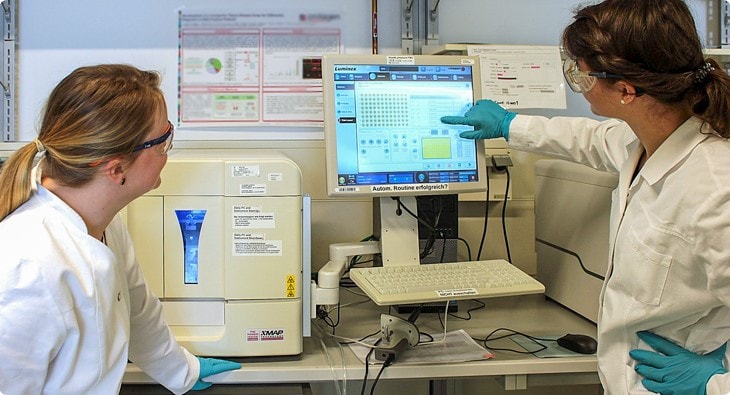
免疫学研究のためのxMAP® テクノロジー
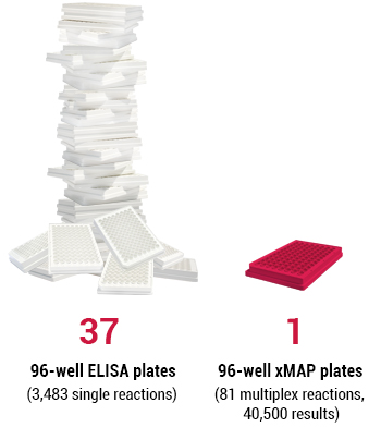
LuminexのxMAP® テクノロジーは、自然免疫プロセス、適応免疫プロセス、細胞シグナル伝達、炎症に関与する複数の可溶性サイトカイン、ケモカイン、免疫グロブリンを測定できます。ビーズベースのxMAP ® テクノロジーは、免疫学研究、創薬、臨床診断で確立されている多くのマルチプレックスイムノアッセイ、遺伝子発現解析、ジェノタイピング、さらに細胞シグナル伝達タンパク質、抗体アイソタイプ、カスタム遺伝子ターゲット測定に使用されています。マルチプレックス解析を用いることで、シングルプレックスのELISAや電気化学発光(ECL)法よりもはるかに多くのデータが得られ、免疫系の制御と機能に関する知識を向上します。
1サンプル、多項目同時解析: xMAP® テクノロジーは複数のターゲットを同時に検出します。
免疫系は非常に複雑であるため、異なる細胞タイプ、様々な抗体、可溶性シグナル伝達因子の相互作用を一括して見ることは困難です。ウェスタンブロットやELISAなどの技術は、ライフサイエンスの研究室では一般的ですが、一度に単一のターゲットしか検出・定量することができません。
xMAP® テクノロジーによるマルチプレックスの利点は、1サンプルから最大500種類のターゲットを同時に検出できるため、複雑な免疫細胞シグナル伝達や血清学に関する洞察を深められることです。

Luminex xMAP® Technology Overview
免疫学の研究 複雑な動き:リンパ球とその機能 免疫系は、異物の侵入に対する身体の反応を仲介するもので、リンパ球を含む複数の種類の細胞で構成されています。リンパ球は、B細胞(抗体を分泌)、細胞傷害性T細胞、ヘルパーT細胞(サイトカインを分泌)、ナチュラルキラー(NK)細胞から構成されています。多くのサイトカインとシグナル伝達タンパク質が、すべてのリンパ球の適切な制御と機能において複雑な役割を担っています。これらのシグナル伝達経路の特性は、リンパ球の相互作用や免疫系の複雑な性質を理解するために不可欠です。 自然免疫系と適応免疫系の細胞は、協調して病原体の攻撃から体を守るために働いています。抗体は、アプリケーションスポットライト: Oncimmune® によるハイスループット自己抗体研究
Oncimmune社の研究者は、既知のヒト抗原の95%以上をカバーする、約9,000の抗原を含む独自の免疫原性タンパク質ライブラリーを開発しました。
免疫チェックポイント阻害剤を投与されている患者の自己抗体プロファイリングのために、数百のサンプルから数千の抗体を検出するには、Oncimmune社はどのようにxMAP® テクノロジー を使用したのでしょうか?ブログでご確認ください。
マルチプレックスで道を拓く: 感染症と治療薬開発
xMAP® テクノロジーは、免疫システムの機能に関する理解を深めるだけでなく、多くの疾患に対するアプローチも変えています。サイトカインや細胞シグナル伝達タンパク質の検出と定量化に加え、抗体のアイソタイプ、リン酸化タンパク質と非リン酸化タンパク質、遊離型と結合型薬剤の解析も可能にします。これらの異なるタイプのマルチプレックスにより、感染症対策や、免疫系と他のシステムとの相互作用に関する知識が大幅に向上し、がん免疫療法の開発のために細胞の機能を操作する方法が提供されるようになりました。
59,000報以上の論文に引用されているxMAP® テクノロジーは、世界で最も利用されているマルチプレックステクノロジーです。
Luminexの文献データベースでは、免疫学研究におけるxMAP® テクノロジーの様々なアプリケーションを検索することができます。
COVID-19と免疫反応: xMAP® テクノロジーを利用したアイソタイピング
COVID-19患者のSARS-CoV-2に対する抗体の初期の測定ではIgGアイソタイプに焦点が当てられていますが、COVID-19に対する身体の免疫反応を完全に理解するにはIgAおよびIgMの反応も重要です。パンデミックの拡大に伴い、患者の免疫状態を把握することは、COVID-19の進行状況を理解する上で非常に重要です。研究者が個人の免疫状態をより正確に把握できるように、血清学的アイソタイプ解析のためのxMAP® 新型コロナウイルスIgG 抗体検出キット (RUO)を拡張する新しいメソッドが設計されました。
このアッセイのアップデートにより、複数のウイルス抗原に対するIgMおよびIgA応答の迅速かつハイスループットな定量化が可能となり、SARS-CoV-2に対するヒトの免疫応答の解明がより一層進むことになります。
柔軟でカスタマイズ可能なプラットフォームでHPVワクチン開発をサポート
ワクチン開発は、従来から複雑で時間のかかるプロセスで、病原体のバリアントとその発症様式、それらが発現する抗原について明確かつ深く理解する必要があります。このような取り組みを成功させるには、関連するすべてのバリアントをタイムリーかつ効率的に検出する高品質のアッセイが必要ですが、従来のELISAが時間と労力を要することは周知のとおりです。ワクチン開発では、ELISAでは必要なスループットを確保することができません。
幸いに、xMAP® ビーズベースのマルチプレックステクノロジーは、遺伝子発現とタンパク質発現の両方のアッセイをサポートする柔軟でカスタマイズ可能なプラットフォームを提供します。xMAP® テクノロジーは、複数のHPV遺伝子型に対する特異的抗体を同時に測定することを可能にし、何千人もの人々の力価測定に使用されています。
これらのHPVワクチン開発に使用されたxMAPアッセイについて、大規模試験の実施方法、特殊な集団を対象とした研究のサポート方法など、より詳しく知りたい方は、Luminexのブログをご覧ください。
種特異的な感染症を識別しながら検査コストを削減
抗原および抗体検査は、集団レベルの傾向や個人の免疫反応などを特徴付けるための重要なデータを提供します。公衆衛生の専門家がアウトブレイク、疾患の傾向、免疫反応を理解するのに役立つ信頼性の高い血清学的検査を開発することを目指し、xMAP® マルチプレックステクノロジーに基づく検査が開発されました。
このプラットフォームのマルチプレックス機能は、1つの抗原だけでなく、複数の抗原を検出できるため、ユーザーは異なる種特異的な病原体を区別できるようになります。したがって、対象者一人一人のPCR検査にサンプルあたりの費用をかけるよりも、サンプルあたりの費用を5分の1に抑え、単一ターゲット検査よりも包括的な結果で同じデータを得ることができます。
マルチプレックスでコストを削減し、より多くのデータを取得する方法をご覧ください。
シングルアッセイでサンプルを節約:従来の免疫原性試験の最適化
免疫原性あるいは生物学的製剤が免疫反応を引き起こす能力を評価することは、創薬における重要なステップです。ワクチン開発では、強い免疫反応は、感染症やワクチンに対する抗体の継続的な発現を意味します。しかし、バイオ医薬品の開発においては、免疫原性の兆候は、抗薬物抗体(ADA)の産生が有害事象につながる可能性や、医薬品の有効性を制限する可能性を示唆しています。
研究者らは、xMAP® テクノロジーを用いて、2つの別々の免疫原性アッセイからマルチプレックスアッセイを設計し、関節リウマチやその他の炎症性疾患の治療によく使用される、免疫原性プロファイルが立証されたTNF阻害剤であるヒュミラ®の投与を受けた患者におけるADAの同定とアイソタイピングを同時に実施しました。
最終的に、マルチプレックステクノロジーは、従来の免疫原性試験の段階的なアプローチを合理化し、作業時間を短縮し、サンプルの量と保全性を維持するのに役立つことが実証されました。xMAP® マルチプレックスアッセイデザインのカスタマイズ、拡張、最適化により、臨床環境における免疫原性評価にどのような価値をもたらすことができるのか、その詳細をご確認ください。
免疫原性試験の結果を犠牲にすることなく、時間とコストを節約することができます。
パートナーから市販されている数千のxMAP® キットが利用可能
ビーズベースのマルチプレックスの汎用性は、Luminexの研究・体外診断用医薬品のパートナーから提供されるサイトカイン、細胞シグナルタンパク質、抗体、遺伝子発現用の既製およびカスタムキットの大規模なポートフォリオによっても明らかです。また、ヒト以外の動物やバクテリアのサンプルにも対応しています。Kit Finderで、パートナーが開発した市販のxMAPアッセイを検索することができます。
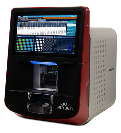
xMAP® システムによるイムノアッセイワークフローの改善
LuminexのxMAP® テクノロジーは、フローサイトメトリーベースの検出とCCDベースのイメージングという2つの検出方式を採用しています。全てのxMAP® システムは正確なデータを提供しますが、様々なラボのマルチプレックスとスループットのニーズにより適したモデルを提供することができます。
あなたのラボに最適なxMAP® システムは?
最適かつ高い汎用性: xMAP INTELLIFLEX™ システムは、デュアルレポーターチャネルを提供します。
最近のxMAP® テクノロジーの進歩には、血清学的アッセイの課題によりよく対応するためのxMAP INTELLIFLEX™ システムの発売があります。低濃度の細胞シグナルタンパク質を測定する場合、2つ目のレポーターチャネルを追加することで、同じタンパク質を標的とする2つの別々の抗体アイソタイプの検出が可能になります。血清学的アッセイに見られるレポーターチャネルが1つであるのに対し、この2つ目のレポーターチャンネルの感度とダイナミックレンジが向上したことにより、サンプル中のアナライトから得られるデータを倍増させることができます。これにより、アッセイの設計、最適化、および検証時に必要な情報を得るためのサンプル量と時間が削減されます。
専門家がLuminexの科学者チームと共同でSARS-CoV-2抗体アッセイを開発した際、何千ものサンプルを用いてアッセイの検証を行った結果、1回の反応で2つの免疫グロブリンのレベルを確実に検出し、同時にSARS-CoV-2中和抗体の生成量を測定することが可能になりました。デュアルレポーターチャネルのユニークな機能により、1回の反応でIgG抗体の検出に加え、IgM抗体の検出にも応用できるようになりました。
マルチタスクとマルチプレックス: xMAP® テクノロジーアッセイの自動化
ラボの自動化とマルチプレックスアッセイを統合することで、特に多検体の解析や高度なマルチプレックスアッセイの開発時に、ワークフロープロセスの効率を大幅に改善することができます。
Luminex のxMAP® テクノロジーは、臨床、産業、ライフサイエンス研究機関において、複数のターゲットの高速かつ反復的なアッセイを必要とするマルチプレックスアッセイ用に設計されています。59,000報以上の論文に見られるように、xMAP® テクノロジーは最も広く使用されているマルチプレックステクノロジーとなっています。
1サンプルで最大500項目のアッセイをマルチプレックスにできるxMAP® プラットフォームは、各ラボのスループットと多項目解析の両方のニーズに応じてアッセイを柔軟にカスタマイズすることが可能です。
さらに、ビーズの均一な分注、ウェル間のキャリーオーバー、液体移動時のビーズロスの最小化などの懸念は、強固な自動化プロトコルで容易に軽減されます。
生産性を高め、エラーを最小限に抑え、研究員をより安全に保つために、研究室にオートメーションを導入しませんか?
免疫学研究のリソースとドキュメント
ホワイトペーパー
その他の資料の検索
ブログ
- Multiplex Technology Optimizes Traditional Immunogenicity Testing
- How Multiplexing Can Support Vaccine Development
- COVID-19 and Characterizing the Immune Response: A Method for Isotyping Using xMAP Technology
- CDC Scientist Describes Multiplex Testing for Malaria at Immunology2022™
- Getting Double the Data from Less Sample: An xMAP INTELLIFLEX® Dual Reporter Assay in Action
関連製品
免疫学研究関連動画
 免疫学の研究 複雑な動き:リンパ球とその機能 免疫系は、異物の侵入に対する身体の反応を仲介するもので、リンパ球を含む複数の種類の細胞で構成されています。リンパ球は、B細胞(抗体を分泌)、細胞傷害性T細胞、ヘルパーT細胞(サイトカインを分泌)、ナチュラルキラー(NK)細胞から構成されています。多くのサイトカインとシグナル伝達タンパク質が、すべてのリンパ球の適切な制御と機能において複雑な役割を担っています。これらのシグナル伝達経路の特性は、リンパ球の相互作用や免疫系の複雑な性質を理解するために不可欠です。 自然免疫系と適応免疫系の細胞は、協調して病原体の攻撃から体を守るために働いています。抗体は、
がん免疫学のバイオマーカーとしての自己抗体
 免疫学の研究 複雑な動き:リンパ球とその機能 免疫系は、異物の侵入に対する身体の反応を仲介するもので、リンパ球を含む複数の種類の細胞で構成されています。リンパ球は、B細胞(抗体を分泌)、細胞傷害性T細胞、ヘルパーT細胞(サイトカインを分泌)、ナチュラルキラー(NK)細胞から構成されています。多くのサイトカインとシグナル伝達タンパク質が、すべてのリンパ球の適切な制御と機能において複雑な役割を担っています。これらのシグナル伝達経路の特性は、リンパ球の相互作用や免疫系の複雑な性質を理解するために不可欠です。 自然免疫系と適応免疫系の細胞は、協調して病原体の攻撃から体を守るために働いています。抗体は、
xMAP INTELLIFLEX システムとイムノアッセイのイノベーション:がん免疫学研究を推進するツール
 免疫学の研究 複雑な動き:リンパ球とその機能 免疫系は、異物の侵入に対する身体の反応を仲介するもので、リンパ球を含む複数の種類の細胞で構成されています。リンパ球は、B細胞(抗体を分泌)、細胞傷害性T細胞、ヘルパーT細胞(サイトカインを分泌)、ナチュラルキラー(NK)細胞から構成されています。多くのサイトカインとシグナル伝達タンパク質が、すべてのリンパ球の適切な制御と機能において複雑な役割を担っています。これらのシグナル伝達経路の特性は、リンパ球の相互作用や免疫系の複雑な性質を理解するために不可欠です。 自然免疫系と適応免疫系の細胞は、協調して病原体の攻撃から体を守るために働いています。抗体は、
COVID-19患者のウイルスエピトープ解析により、交差反応性と重症度の相関が判明
 免疫学の研究 複雑な動き:リンパ球とその機能 免疫系は、異物の侵入に対する身体の反応を仲介するもので、リンパ球を含む複数の種類の細胞で構成されています。リンパ球は、B細胞(抗体を分泌)、細胞傷害性T細胞、ヘルパーT細胞(サイトカインを分泌)、ナチュラルキラー(NK)細胞から構成されています。多くのサイトカインとシグナル伝達タンパク質が、すべてのリンパ球の適切な制御と機能において複雑な役割を担っています。これらのシグナル伝達経路の特性は、リンパ球の相互作用や免疫系の複雑な性質を理解するために不可欠です。 自然免疫系と適応免疫系の細胞は、協調して病原体の攻撃から体を守るために働いています。抗体は、
真菌によるアレルギー性気道炎症における免疫防御反応と免疫病原性反応
 免疫学の研究 複雑な動き:リンパ球とその機能 免疫系は、異物の侵入に対する身体の反応を仲介するもので、リンパ球を含む複数の種類の細胞で構成されています。リンパ球は、B細胞(抗体を分泌)、細胞傷害性T細胞、ヘルパーT細胞(サイトカインを分泌)、ナチュラルキラー(NK)細胞から構成されています。多くのサイトカインとシグナル伝達タンパク質が、すべてのリンパ球の適切な制御と機能において複雑な役割を担っています。これらのシグナル伝達経路の特性は、リンパ球の相互作用や免疫系の複雑な性質を理解するために不可欠です。 自然免疫系と適応免疫系の細胞は、協調して病原体の攻撃から体を守るために働いています。抗体は、
xMAP INTELLIFLEX システムを使ったCOVID-19ワクチン有効性評価アッセイのデザイン
 免疫学の研究 複雑な動き:リンパ球とその機能 免疫系は、異物の侵入に対する身体の反応を仲介するもので、リンパ球を含む複数の種類の細胞で構成されています。リンパ球は、B細胞(抗体を分泌)、細胞傷害性T細胞、ヘルパーT細胞(サイトカインを分泌)、ナチュラルキラー(NK)細胞から構成されています。多くのサイトカインとシグナル伝達タンパク質が、すべてのリンパ球の適切な制御と機能において複雑な役割を担っています。これらのシグナル伝達経路の特性は、リンパ球の相互作用や免疫系の複雑な性質を理解するために不可欠です。 自然免疫系と適応免疫系の細胞は、協調して病原体の攻撃から体を守るために働いています。抗体は、
xMAPアッセイの自動化
本製品は研究用の製品です。体外診断用には使用できません。