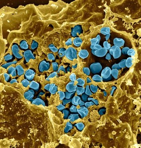
Macrophage infected with Francisella tularensis bacteria. Photo credit: National Institute of Allergy and Infectious Disease (NIAID). This work is licensed under a Creative Commons Attribution 2.0 Generic License.
Researchers recently studied Francisella tularensis, a highly virulent bacteria which results in tularemia in mammals (including humans). The bacteria takes up residence in macrophages, proliferating in the cytosol. To do this most effectively, F. tularensis modifies the host cell death cycle. Its ability to synchronize its proliferation in the cytosol with cell death is key to its virulence and ability to bypass the host’s immunological response. This mechanism seems to be primarily apoptotic, with some characteristics of pyroptosis also present. This study hopes to provide a deeper understanding of this bacteria’s poorly understood mechanism.
To do this, the authors of “Importance of PdpC, IglC, IglI, and IglG for modulation of a host cell death pathway induced by Francisella tularensis LVS”1 utilized the Luminex® multiplexing system to test macrophages infected with the live vaccine strain (LVS) and compared its effects to those of several deletion mutations using mouse specific anti-caspase-9 antibody to detect pro-caspase-9 and cleaved caspase-9, which indicate mitochondrial membrane destabilization. Several other measures of modulation were also studied.
For cytokine analysis, the Luminex 100/200™ and Bio-Plex Pro™ Mouse cytokine 23-plex kit were used for multiplexing, which meant getting the most out of a limited supply of mouse samples. They “performed a comprehensive multiplex analysis of the secretion of cytokines and chemokines from J774 cells infected with either of the four mutants or the LVS strain.”1
The study concluded that the secreted cytokines indicate the ΔpdpC mutant-infected cells showed the most similar phenotype to the LVS-infected cells. Our present findings further corroborate the unusual phenotype of the ΔpdpC mutant since it was found to be distinct to all other investigated mutants, and infection led to marked 16 mitochondrial damage, caspase-3 cleavage, expression of PS, and DNA fragmentation, although with delayed kinetics compared to LVS. Moreover, the secreted cytokine pattern was very similar to that of LVS-infected cell. Their findings contribute to a better understanding of the cytopathogenic effects of F. tularensis infection and its mechanisms for signaling cell death in the host.
The use of Luminex multiplexing cytokine analysis meant that many more tests could be run on their samples with limited available amounts of sample. This level of efficient testing was therefore invaluable to completing this research. Multiplexing was the most cost-effective, accurate means of analysis of their available samples.
Learn more about Luminex multiplexing instruments.
Looking for immunoassay kits from our partners? Check out xMAP® Kit Finder.
References
- Lindgren M, Eneslätt K, Bröms JE, Sjöstedt A. Importance of PdpC, IglC, IglI, and IglG for modulation of a host cell death pathway induced by Francisella tularensis. Infection and Immunity. 2013 Jun;81(6):2076-84. Doi: 10.1128/IAI.00275-13. Epub 2013 Mar 25. Available from: http://www.ncbi.nlm.nih.gov/pubmed/23529623.