Multiplex immunoassays compared with brain cancer biomarker detection

Accurately staging any cancer can be challenging, but assessing glioblastomas hidden in the brain is even more difficult. Scientists are working hard to discover and validate blood-based biomarkers that could provide useful prognostic and diagnostic information for these malignancies while overcoming the major accessibility issue associated with brain tumors. Cytokines are especially promising since they are released as part of the natural immune response to glioblastomas.
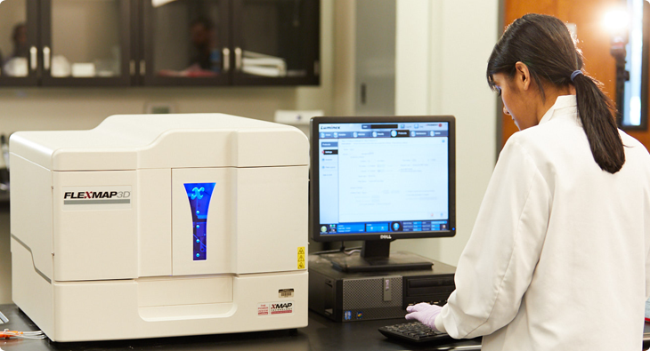
Recently, a team of scientists at Washington University School of Medicine aimed to evaluate the performance of two different multiplex immunoassay techniques for cytokine profiling in plasma samples from patients with brain tumors and healthy volunteers. They compared an assay on the Luminex FLEXMAP 3D® system to planar electrochemiluminescence (ECL) kits from another vendor, and both techniques were performed in 96-well plates.
Comparing two leading immunoassay platforms
The Luminex assay covered 21 cytokines in a single reaction, while it took three separate ECL kits to cover 20 cytokines. The team evaluated performance based on the 19 cytokines common to both methods, using the same set of samples for each: 27 samples from glioblastoma patients, 17 samples from subjects with cancer metastasized to the brain, and 11 samples from healthy controls.
With the FLEXMAP 3D system, all 19 cytokines were quantified in all 55 samples. The ECL kits, though, struggled to perform as well. While calibration curves suggested a lower limit of detection, the kits failed to detect seven of the 19 cytokines in more than 25% of samples. One cytokine, IL-21, could not be detected in any sample using the ECL kits. Collectively, these data indicate that the Luminex technology offers superior performance with real-world samples for rapidly quantifying many cytokines.
In addition, the team analyzed the operator time needed for each approach. They found that the Luminex technique took 1 hour and 38 minutes of hands-on time, while the ECL kits took 2 hours and 42 minutes of hands-on time. The longer time was associated with having to run multiple ECL plates to cover the target number of cytokines.
Overall, the team determined that multiplex assays have advantages over singleplex colorimetric ELISAs, as they streamline cytokine profiling while simultaneously decreasing cost and time in motion.
If you’re interested in learning more about how xMAP Technology can streamline your workflow and expand your research, we’ve developed a free cost comparison tool that demonstrates how much time and money you can save by entering your custom lab parameters.
For Research Use Only. Not for use in diagnostic procedures.
Related Content
- Multiplex Technology Optimizes Traditional Immunogenicity Testing [Blog]
- xMAP® Cookbook to Design Your Own Assays [Download]
- Meet xMAP® INTELLIFLEX! A First Look at the Newest xMAP® Technology Platform [Blog]
- Browse 1,200+ Partner Kits with xMAP® Kit Finder [Online Tool]
- The Versatility of Multiplex Antibody Titer Assays for COVID-19 and Beyond [Blog]
